
TALE homeodomain transcription factors.Keratinocytes are the only cells in the body with the entire vitamin D metabolic pathway from vitamin D production to catabolism and vitamin D receptor expression. Vitamin D 3 (cholecalciferol) regulates keratinocyte proliferation and differentiation mostly by modulating calcium concentrations and regulating the expression of genes involved in keratinocyte differentiation.Moreover, it has been suggested that an extracellular calcium-sensing receptor (CaSR) also contributes to the rise in intracellular calcium concentration. Part of that intracellular calcium increase comes from calcium released from intracellular stores and another part comes from transmembrane calcium influx, through both calcium-sensitive chloride channels and voltage-independent cation channels permeable to calcium.
Translucent cells containing keratin called free#
Those elevations of extracellular calcium concentrations induces an increase in intracellular free calcium concentrations in keratinocytes. Calcium concentration in the stratum corneum is very high in part because those relatively dry cells are not able to dissolve the ions.

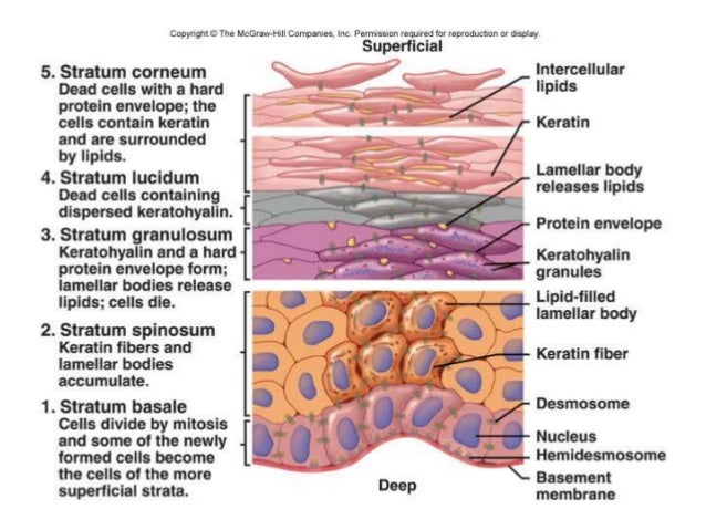
A calcium gradient, with the lowest concentration in the stratum basale and increasing concentrations until the outer stratum granulosum, where it reaches its maximum.įactors promoting keratinocyte differentiation are: In humans, it is estimated that keratinocytes turn over from stem cells to desquamation every 40–56 days, whereas in mice the estimated turnover time is 8–10 days. Corneocytes will eventually be shed off through desquamation as new ones come in.Īt each stage of differentiation, keratinocytes express specific keratins, such as keratin 1, keratin 5, keratin 10, and keratin 14, but also other markers such as involucrin, loricrin, transglutaminase, filaggrin, and caspase 14. ĭuring this differentiation process, keratinocytes permanently withdraw from the cell cycle, initiate expression of epidermal differentiation markers, and move suprabasally as they become part of the stratum spinosum, stratum granulosum, and eventually corneocytes in the stratum corneum.Ĭorneocytes are keratinocytes that have completed their differentiation program and have lost their nucleus and cytoplasmic organelles. Those stem cells and their differentiated progeny are organized into columns named epidermal proliferation units. Some of the transit amplifying cells continue to proliferate then commit to differentiate and migrate towards the surface of the epidermis. Epidermal stem cells divide in a random manner yielding either more stem cells or transit amplifying cells. Cell differentiation Įpidermal stem cells reside in the lower part of the epidermis (stratum basale) and are attached to the basement membrane through hemidesmosomes. The fully cornified keratinocytes that form the outermost layer are constantly shed off and replaced by new cells. Keratinization is part of the physical barrier formation ( cornification), in which the keratinocytes produce more and more keratin and undergo terminal differentiation. Structure Ī number of structural proteins ( filaggrin, keratin), enzymes ( proteases), lipids, and antimicrobial peptides ( defensins) contribute to maintain the important barrier function of the skin.

Pathogens invading the upper layers of the epidermis can cause keratinocytes to produce proinflammatory mediators, particularly chemokines such as CXCL10 and CCL2 (MCP-1) which attract monocytes, natural killer cells, T-lymphocytes, and dendritic cells to the site of pathogen invasion. The primary function of keratinocytes is the formation of a barrier against environmental damage by heat, UV radiation, water loss, pathogenic bacteria, fungi, parasites, and viruses. Keratinocytes differentiate from epidermal stem cells in the lower part of the epidermis and migrate towards the surface, finally becoming corneocytes and eventually be shed off, which happens every 40 to 56 days in humans. Keratinocytes form a barrier against environmental damage by heat, UV radiation, water loss, pathogenic bacteria, fungi, parasites, and viruses.Ī number of structural proteins, enzymes, lipids, and antimicrobial peptides contribute to maintain the important barrier function of the skin.
Translucent cells containing keratin called skin#
Basal cells in the basal layer ( stratum basale) of the skin are sometimes referred to as basal keratinocytes. In humans, they constitute 90% of epidermal skin cells. Keratinocytes are the primary type of cell found in the epidermis, the outermost layer of the skin. Primary type of cell found in the epidermis Micrograph of keratinocytes, basal cells and melanocytes in the epidermis Keratinocytes (stained green) in the skin of a mouse


 0 kommentar(er)
